6 years male child was diagnosed clinically as asthma and put on controller medication in the form of Inhalational Budesonide for dialy use with inhalational salbutamol as and when required
After the initial control for nearly 2 months the child came to OPD with shortness of breath sometimes in last 2 weeks and dyspnea on exertion
Clinical examination was done which revealed no respiratory distress with bilateral wheeze on chest auscultation
Spirometry (PFT) was done which was normal .
Child was sent home after increasing the dose of Budesonide as the technique of inhalation and adherence was perfect and no comorbidity or recent allergen exposure was detected
Child came back after 2 weeks with no improvement.
This time it was detected on chest auscultation that the air entry was diminished over left infraclavicular region.
Chest X-Ray was done which revealed opacity in the left upper lobe.
CECT chest was done to clearly define the opacity ,which revealed large cystic lesion with arterial supply from pulmonary circulation
The Diagnosis was –CPAM
Case discussion – In the diagnosis and management of asthma , congenital lung malformations should be kept a possibility specially in early childhood asthma.
In case of no adequate response to standard asthma treatment, radio imaging should be done to rule out other lung lesions
Congenital pulmonary airway malformations (CPAM)occur during defect in lung morphogenesis in embryonic stages.Different types of CPAM occurs at different stages of defective morphogenesis.
CPAM is classified into 5 types
-Type 0- is also known as acinar dysplasia and it is rare
Type 1- is cystic lesions one or more with more than 2 cm cystic diameter
Type 2- is multiple cystic lesions with cystic diameter of less than 2 cm-this is the most common type seen
Type 3-is solid lesions
Type 4- is multi loculated cystic lesions located peripherally near the pleura
Type 4 may be complicated by Pneumothorax and rarely converts into Pleuropulmonary blastoma ,a malignant lung condition
CPAM is detected in utero by USG in second trimester of pregnancy. If it is is large enough endangering the fetal life , intrauterine aspiration ,corticosteroid or surgical resection are indicated
After birth, if the infant is symptomatic or if CPAM occupies more than 20 % volume of hemithorax ,surgical resection should be done. Surgical resection is also indicated in type 4 CPAM to prevent Pleuropulmonary Blastoma specially in presence of POSITIVE family history of DICER 1 gene .
Other indication for surgery -to prevent repeated pulmonary infection, bilateral lesions, multifocal cysts.
An association of Type 1 CPAM has been found with Bronchoalveolar carcinoma
References: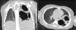
Maneenil G, Ruangnapa K, Thatrimontrichai A, Janjindamai W, Dissaneevate S, Anantaseree W, Suntornlohanakul S. Clinical presentation and outcome in congenital pulmonary malformation: 25 year retrospective study in Thailand. Pediatrics international : official journal of the Japan Pediatric Society. 2019 Aug:61(8):812-816. doi: 10.1111/ped.13934. Epub 2019 Jul 2 [PubMed PMID: 31264305]
Parikh DH, Rasiah SV. Congenital lung lesions: Postnatal management and outcome. Seminars in pediatric surgery. 2015 Aug:24(4):160-7. doi: 10.1053/j.sempedsurg.2015.01.013. Epub 2015 Feb 3 [PubMed PMID: 26051048]
For more knowledge of Pediatric Pulmonology ,join Fellowship in Pediatric Pulmonology with Bronchoscopy and Sleep Study under aegis of RespiratoRY Foundation of India-click here to apply for the course-https://respifoundation.com